
Donkey Secondary Antibodies


Biotium Spectrin beta III (SPTBN2) (SPTBN2/1582), CF647 conjugate, 0.1mg/mL
Spectrin is an actin binding protein that is a major component of the plasma membrane skeleton. Spectrins function as membrane organizers and stabilizers by forming dimers, tetramers and higher polymers. Vertebrate spectrins have two alpha-subunits (alpha-I/alpha-II), four beta-subunits (beta-I-beta-IV) and a beta-H subunit creating diversity and specialization of function. Spectrin alpha and spectrin beta are present in erythrocytes, whereas spectrin alpha II (also designated fodrin alpha) and spectrin beta I (also designated fodrin beta) are present in other somatic cells. The spectrin tetramers in erythrocytes act as barriers to lateral diffusion, but spectrin dimers seem to lack this function. Spectrin beta III is highly homologous to both spectrin beta I and spectrin beta II. Spectrin beta III is highly expressed in brain, kidney, pancreas and liver, and at lower levels in lung and placenta. Spectrin beta 3 is primarily expressed in nervous tissues with highest expressio

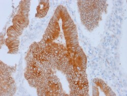
Biotium Aurora B (Proliferation Marker)(AURKB/1592), CF647 conjugate, 0.1mg/mL
This antibody recognizes a protein of 39 kDa, which is identified as Aurora kinase B (or Aurora B). Aurora B is a serine/threonine protein kinase and component of the chromosomal passenger complex (CPC), a complex that acts as a key regulator of mitosis. It is important for accurate chromosome segregation, cytokinesis, protein localization to the centromere and kinetochore, correct microtubule-kinetochore attachment, and regulation of the mitotic checkpoint. Aurora B forms a tight complex with inner centrosome protein and survivin. Inactivation of any of these proteins causes similar defects in chromosome segregation. A significant overexpression of Aurora B has been found in a variety of human tumors including non-small cell lung carcinoma, astrocytoma, seminoma and carcinomas of the colon, prostate, endometrium and thyroid. The expression level of Aurora B is associated with cell proliferation and prognosis in these tumors. Primary antibodies are available purified, or with
Biotium Connexin 32 (Gap Junction Protein) (R5.21C), CF647 conjugate, 0.1mg/mL
This antibody recognizes a protein of 27-32 kDa, identified as Connexin 32. The connexin family of proteins forms hexameric complexes called connexons that facilitate movement of low molecular weight proteins between cells via gap junctions. Connexin proteins share a common topology of four transmembrane alphalpha-helicaldomains, two extracellular loops, a cytoplasmic loop and cytoplasmic N- and C-termini. Many of the key functional differences arise from specific amino-acid substitutions in the most highly conserved domains, the transmembrane and extracellular regions. Each of the approximately 20-connexin isoforms produces channels with distinct permeability and electrical and chemical sensitivities; therefore, one connexin usually cannot fully substitute for another.Primary antibodies are available purified, or with a selection of fluorescent CF Dyes and other labels. CF Dyes offer exceptional brightness and photostability. Note: Conjugates of blue fluorescent dyes like
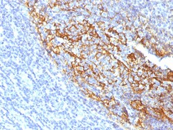
Biotium CD35 / CR1 (Follicular Dendritic Cell Marker)(To5), CF647 conjugate, 0.1mg/mL
Recognizes a protein of 210-220 kDa, which is identified as the complement receptor 1 (CR1)/CD35. This MAb does not block CR1 activity. It is highly specific to CR1 and shows no cross-reaction with CR2. The primary function of CR1 is to serve as the cellular receptor for C3b and C4b, the most important components of the complement system leading to clearance of foreign macromolecules. The Knops blood group system is a system of antigens located on this protein. Follicular dendritic cells (FDC) are restricted to the B-cell regions of secondary lymphoid follicles. They are CD21 /CD35 /CD1a-. This MAb labels follicular dendritic cells and follicular dendritic cell sarcoma. Primary antibodies are available purified, or with a selection of fluorescent CF Dyes and other labels. CF Dyes offer exceptional brightness and photostability. Note: Conjugates of blue fluorescent dyes like CF405S and CF405M are not recommended for detecting low abundance targets, because blue dyes have
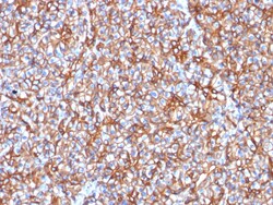
Biotium Spectrin beta III (SPTBN2) (SPTBN2/1778), CF647 conjugate, 0.1mg/mL
Spectrin is an actin binding protein that is a major component of the plasma membrane skeleton. Spectrins function as membrane organizers and stabilizers by forming dimers, tetramers and higher polymers. Vertebrate spectrins have two alpha-subunits (alpha-I/alpha-II), four beta-subunits (beta-I-beta-IV) and a beta-H subunit creating diversity and specialization of function. Spectrin α and spectrinβ are present in erythrocytes, whereas spectrin α II (also designated fodrin α) and spectrinβ I (also designated fodrinβ) are present in other somatic cells. The spectrin tetramers in erythrocytes act as barriers to lateral diffusion, but spectrin dimers seem to lack this function. Spectrinβ III is highly homologous to both spectrinβ I and spectrinβ II. Spectrinβ III is highly expressed in brain, kidney, pancreas and liver, and at lower levels in lung and placenta. Spectrin beta 3 is primarily expressed in nervous tissues with high
Biotium Factor XIIIa (Coagulation Factor XIIIA Chain) (F13A1/1683), CF647 conjugate, 0.1mg/mL
This antibody recognizes a protein of 83 kDa, which is identified as Factor XIIIa. It has been identified in platelets, megakaryocytes, and fibroblast-like mesenchymal or histiocytic cells in the placenta, uterus, and prostate, monocytes and macrophages and dermal dendritic cells. Anti-factor XIIIa has been found to be useful in differentiating between dermatofibroma (almost all cases are positive), dermatofibrosarcoma protuberans (-/ ) and desmoplastic malignant melanoma (-). Anti-factor XIIIa positivity is also seen in capillary hemagioblastoma, hemangioendothelioma, hemangiopericytoma, xanthogranuloma, xanthoma, hepatocellular carcinoma, glomus tumor, and meningioma.Primary antibodies are available purified, or with a selection of fluorescent CF Dyes and other labels. CF Dyes offer exceptional brightness and photostability. Note: Conjugates of blue fluorescent dyes like CF405S and CF405M are not recommended for detecting low abundance targets, because blue dyes have lowe
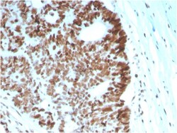
Biotium MSH6 (DNA Mismatch Repair Protein)(MSH6/2927), CF647 conjugate, 0.1mg/mL
The finding that mutations in DNA mismatch repair genes are associated with hereditary nonpolyposis colorectal cancer (HNPCC) has resulted in considerable interest in the understanding of the mechanism of DNA mismatch repair. Initially, inherited mutations in the MSH2 and MLH1 homologs of the bacterial DNA mismatch repair genes mutS and mutL were demonstrated at high frequency in HNPCC and were shown to be associated with microsatellite instability. A member of the mismatch repair family, GTBP (also designated MSH6), is an MSH2-related protein that binds to DNA containing G/T mismatches. Findings suggest that the mismatch-binding factor in human cells is composed of a heterodimer of GTBP and MSH2. Primary antibodies are available purified, or with a selection of fluorescent CF Dyes and other labels. CF Dyes offer exceptional brightness and photostability. Note: Conjugates of blue fluorescent dyes like CF405S and CF405M are not recommended for detecting low abundance targ
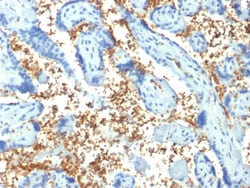
Biotium Spectrin Alpha 1 (Erythrocyte Marker) (SPTA1/1810), CF647 conjugate, 0.1mg/mL
Spectrin is an actin crosslinking and molecular scaffold protein that links the plasma membrane to the actin cytoskeleton, and functions in the determination of cell shape, arrangement of transmembrane proteins, and organization of organelles. Spectrins function as membrane organizers and stabilizers, composed of non-homologous α andβ chains, which aggregate side-to-side in an anti-parallel fashion to form dimers, tetramers and higher polymers. Spectrin α I and spectrinβ I are present in erythrocytes, whereas spectrin α II (also designated fodrin α) and spectrinβ II (also designated fodrinβ) are present in other somatic cells. The spectrin tetramers in erythrocytes act as barriers to lateral diffusion, but spectrin dimers seem to lack this function.Primary antibodies are available purified, or with a selection of fluorescent CF Dyes and other labels. CF Dyes offer exceptional brightness and photostability. Note: Conjugates of blue fluorescent dyes like CF405S
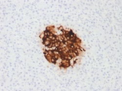
Biotium Insulin / IRDN (beta-Cell & Insulinoma Marker) (rIRDN/805), CF647 conjugate, 0.1mg/mL
This antibody recognizes a polypeptide which is identified as insulin, a 51-amino acid polypeptide composed of A and B chains connected through the C-peptide. Proinsulin, which has very little biological activity, is cleaved by proteases within its cell of origin into the insulin molecule and the C-terminal basic residue. Insulin enhances membrane transport of glucose, amino acids, and certain ions. It also promotes glycogen storage, formation of triglycerides, and synthesis of proteins and nucleic acids. Deficiency of insulin results in diabetes mellitus. The main storage site for insulin is the pancreatic islets. Antibodies to insulin are important as beta-cell and insulinoma markers.Primary antibodies are available purified, or with a selection of fluorescent CF Dyes and other labels. CF Dyes offer exceptional brightness and photostability. Note: Conjugates of blue fluorescent dyes like CF405S and CF405M are not recommended for detecting low abundance targets, because
Biotium MMP3 (Marker of Metastasis and Rheumatoid Arthritis)(MMP3/1994R), CF647 conjugate, 0.1mg/mL
The matrix metalloproteinases (MMP) are a family of peptidase enzymes responsible for the degradation of extracellular matrix components, including collagen, gelatin, fibronectin, laminin and proteoglycan. Transcription of MMP genes is differentially activated by phorbol ester, lipopolysaccharide (LPS) or staphylococcal enterotoxin B (SEB). MMP catalysis requires both calcium and zinc. MMP-3, MMP-10 and MMP-11 (also designated stromelysin-1, 2 and 3, respectively) activate procollagenase. MMP-3 activation of procollagenase can occur via two pathways. Direct activation by MMP-3 is slow and activation by MMP-3 in conjunction with tissue or plasma proteinases is rapid. MMP-10 is expressed in small intestine, and at lower levels in lung and heart. MMP-11 is specifically expressed in stromal cells of breast carcinomas and contributes to epithelial cell malignancies. Primary antibodies are available purified, or with a selection of fluorescent CF Dyes and other labels. CF Dyes of

Biotium CA19-9/Sialyl Lewisa (GI Tumor Marker) (CA19.9/1390R), CF647 conjugate, 0.1mg/mL
CA19-9, a carbohydrate epitope expressed on a high MW (>400 kDa) mucin glycoprotein, is a sialyl Lewisa structure which is synthesized from type 1 blood group precursor chains and is present in individuals expressing the Lewisa and/or Lewisb blood group antigens. In normal tissues, sialyl Lewisa antigen is present in ductal epithelium of the breast, kidney, salivary gland, and sweat glands. Its expression is greatly enhanced in serum as well as in the majority of tumor cells in gastrointestinal (GI) carcinomas, including adenocarcinomas of the stomach, intestine, and pancreas. Preoperative elevated CA19-9 levels in patients with stage I pancreatic carcinoma decrease to normal values following surgery. When used serially, CA19-9 can predict recurrence of disease prior to radiographic or clinical findings. This MAb is excellent for staining of formalin-fixed, paraffin-embedded tissues.Primary antibodies are available purified, or with a selection of fluorescent CF Dyes an
Biotium CD2(UMCD2), CF647 conjugate, 0.1mg/mL
CD2 interacts through its amino-terminal domain with the extracellular domain of CD58 (also designated CD2 ligand) to mediate cell adhesion. CD2/CD58 binding can enhance antigen-specific T cell activation. CD2 is a transmembrane glycoprotein that is expressed on peripheral blood T lymphocytes, NK cells and thymocytes. CD58 is a heavily glycosylated protein with a broad tissue distribution in hematopoietic and other cells, including endothelium. Interaction between CD2 and its counter receptor LFA3 (CD58) on opposing cells optimizes immune system recognition, thereby facilitating communication between helper T lymphocytes and antigen-presenting cells, as well as between cytolytic effectors and target cells.Primary antibodies are available purified, or with a selection of fluorescent CF Dyes and other labels. CF Dyes offer exceptional brightness and photostability. Note: Conjugates of blue fluorescent dyes like CF405S and CF405M are not recommended for detecting low abunda
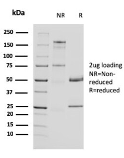
Biotium Uroplakin 1A (Urothelial Differentiation Marker)(UPK1A/2924), CF647 conjugate, 0.1mg/mL
The protein encoded by this gene is a member of the transmembrane 4 superfamily, also known as the tetraspanin family. Most of these members are cell-surface proteins that are characterized by the presence of four hydrophobic domains. The proteins mediate signal transduction events that play a role in the regulation of cell development, activation, growth and motility. This encoded protein is found in the asymmetrical unit membrane (AUM) where it can complex with other transmembrane 4 superfamily proteins. It may play a role in normal bladder epithelial physiology, possibly in regulating membrane permeability of superficial umbrella cells or in stabilizing the apical membrane through AUM/cytoskeletal interactions. The protein may also play a role in tumor suppression. Primary antibodies are available purified, or with a selection of fluorescent CF Dyes and other labels. CF Dyes offer exceptional brightness and photostability. Note: Conjugates of blue fluorescent dyes like
Biotium Ksp-Cadherin / CDH16(CDH16/1071), CF647 conjugate, 0.1mg/mL
This MAb recognizes a protein of 130 kDa, identified as Ksp-cadherin. Cadherins form a superfamily of related glycoproteins that mediate calcium-dependent cell adhesion and transmit signals from the extracellular matrix to the cytoplasm. Cadherins have been implicated in embryogenesis, tissue morphogenesis, tissue structure maintenance, cell polarization, neoplastic invasiveness and metastasis, and membrane transport. It is suggested that Ksp-cadherin is a marker for terminal differentiation of the basolateral membranes of renal tubular epithelial cells. Within the kidney, Ksp-Cadherin is found exclusively in the basolateral membrane of renal tubular epithelial cells and collecting duct cells, and not in glomeruli, renal interstitial cells, or blood vessels. Ksp-Cadherin has been suggested to distinguish Chromophobe Renal-Cell Carcinoma from Oncocytoma.Primary antibodies are available purified, or with a selection of fluorescent CF Dyes and other labels. CF Dyes offer except
Biotium CD70(BU69), CF647 conjugate, 0.1mg/mL
This antibody recognizes a protein of 30 kDa, identified as CD70. It is a cytokine that belongs to the tumor necrosis factor (TNF) ligand family. This cytokine is a ligand for TNFRSF27/CD27. It is a surface antigen on activated, but not on resting, T- and B-lymphocytes. It induces proliferation of co-stimulated T cells, enhances the generation of cytolytic T cells, and contributes to T cell activation. This cytokine is also reported to play a role in regulating B-cell activation, cytotoxic function of natural killer cells, and immunoglobulin synthesis. This MAb blocks the interaction between CD27 and CD70, and has been shown toinhibit T cell proliferation induced by dendritic cells.Primary antibodies are available purified, or with a selection of fluorescent CF Dyes and other labels. CF Dyes offer exceptional brightness and photostability. Note: Conjugates of blue fluorescent dyes like CF405S and CF405M are not recommended for detecting low abundance targets, because blu